Glycerol Free or Lyophilized T4 gp32 Protein is useful for many applications including PCR and RPA (Recombinase Polymerase Amplification).
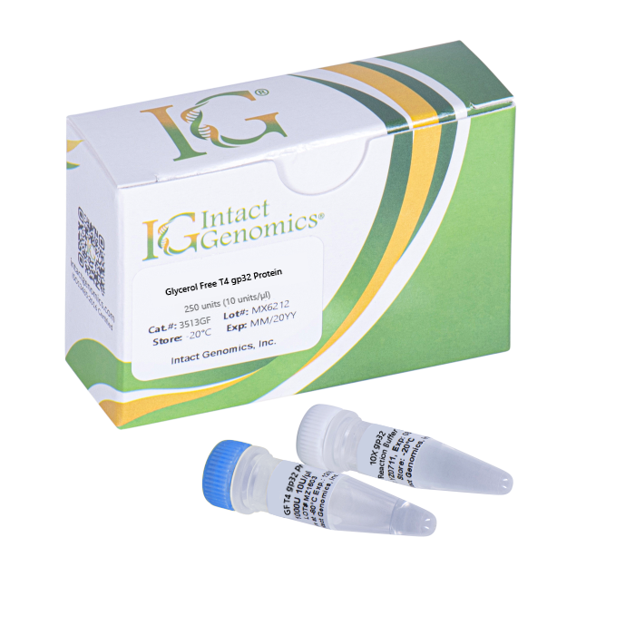
T4 gp32 Protein, Glycerol-free or Lyophilized
Price range: $170.00 through $570.00
Description
T4 gp32 Protein is a single-stranded DNA binding protein required for T4 DNA replication, recombination, and repair (1, 2). It improves the efficiency of reverse transcriptase (RT) during RT-PCR (3), enhances T4 DNA polymerase activity (4), as well as increases the yield of PCR products (5).
Note:
Glycerol acts as a cryoprotectant and protein stabilizer when added to the storage buffer of proteins and enzymes. However, in certain circumstances, it is preferred to omit glycerol from the buffer. This includes instances where the presence of glycerol may interfere, such as lyophilization, high-throughput instruments with sensitive fluidics or primary cell cultures.
For our glycerol free products, it is recommended to use immediately upon thaw as without glycerol there will be significant activity loss with freeze/thaw cycles.
Protein Purity
The physical purity of this enzyme is ≥99% as assessed by SDS-PAGE with Coomassie® blue staining (Fig. 1).
Product Source
- coli BL21 (DE3) strain expressing T4 gp32 gene.
Product Includes
- Glycerol Free T4 gp32 protein
- 10X gp32 reaction buffer
1x GP 32 reaction buffer composition
20 mM Tris-acetate
100 mM Potassium acetate
10 mM Magnesium acetate
1 mM DTT
pH 7.8 @ 25ºC
Storage Buffer
50 mM Tris-HCl
50 mM KCl
1 mM DTT
0.1 mM EDTA
pH 7.5 @ 25ºC
Storage Temperature
–20ºC
Heat Inactivation
65°C for 20 min
Quality Control assays
T4 gp32 protein is free from detectable nuclease activities.
References
- Chase, J. W. and Williams, K. R. (1986) Ann. Rev. Biochem. 55, 103-136
- Sinha, N. K. and Snustad, D. P. (1971) J. Mol. Biol. 62, 267-271.
- Baugh, L.R. et al. (2001). Nucl. Acids Res. 29, e29.
- Topal, M.D. and Sinha, N.K. (1983). J. Biol. Chem. 258, 12274-12279.
- Schwartz, K. et al. (1990). Nucl. Acids Res. 18, 1079.
3513GF, 3516GF, 3519GF, 3513Lyo, 3516Lyo, 3519Lyo, 3519 Lyo
Additional information
| µg | 250µg (10µg/µl), Glycerol-free, 500µg (10µg/µl), Glycerol-free, 1000µg (10µg/µl), Glycerol-free, 250µg (10µg/µl), Lyophilized, 500µg (10µg/µl), Lyophilized, 1000µg (10µg/µl), Lyophilized |
|---|
Intact Genomics Concentration Determination Method
IG uses orthogonal, 3-part approaches to determine the enzyme concentration to provide you with consistent and reliable enzymes for your needs. The quantity of a protein sample is assessed using densitometry with polyacrylamide gel electrophoresis (PAGE), UV absorbance spectra of native protein, and using a protein standard assay such as bicinchoninic acid assay (BCA) using bovine serum albumin (BSA) as a standard (Figure 1).
Why does IG use all three approaches?
1. Each method above has limitations. The limitations include experimental noise, accuracy, and susceptibility to buffer and/or enzyme conditions.
2. Each enzyme has unique physical properties that make a single approach to analyzing proteins a challenge. Each enzyme has a different protein sequence, different requirements to be stable in solution, and different requirements to retain its maximal activity. These differences can interfere with or convolute results, especially when compared to other enzymes. When used together, however, each method provides the scientist with independent measures of both enzyme and buffer purity and quality.
 Figure 1: Enzyme quantitation methods used by IG. A) SDS-polyacrylamide gel electrophoresis. Ladder in 1st lane, 2 µg BSA (~67 kDa) as a standard in lanes 1-3, and IG enzymes (~40 kDa) in lanes 4-6. The yellow boxes are the areas evaluated by densitometry. The integrated band intensities of IG enzymes are compared with integrated band intensities from BSA to assay concentration. B) UV spectrum of a clean IG enzyme with protein peaks at 230 nm and at 280 nm. An extinction coefficient at 280 nm is typically used to quantify protein using these spectra with buffer subtraction at 330 nm. C) BCA standard curve for BSA. The curve is used to calculate an IG enzyme concentration using BSA as the standard.