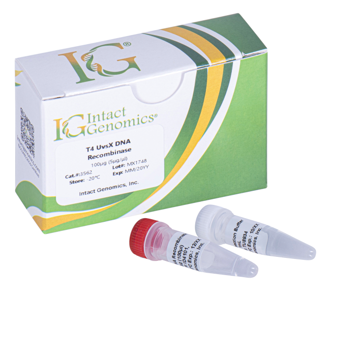
T4 UvsX DNA Recombinase is useful for Recombinase Polymerase Amplification (RPA) and other applications.
T4 UvsX DNA Recombinase
Price range: $235.00 through $1,185.00
Description
Homologous recombination is important for the error-free repair of DNA double-strand breaks and for replication fork restart. Recombinases of the RecA/RAD51 family perform the central catalytic role in this process. UvsX recombinase is the RecA/Rad51 ortholog of bacteriophage T4. T4 UvsX DNA Recombinase and other recombinases form presynaptic filaments on ssDNA that are activated to search for homology in dsDNA and to perform DNA strand exchange (1-3).
Protein Purity
The physical purity of this enzyme is ≥98% as assessed by SDS-PAGE with Coomassie® blue staining (Fig. 1).
Product Source
- Ecoli BL21 (DE3) strain expressing T4 UvsX gene.
Product Components
- UvsX Recombinase
- 10X UvsX Recombinase Reaction Buffer
1x UvsX Recombinase Reaction Buffer Composition
20 mM Tris-acetate pH 7.8
100 mM Potassium acetate
10 mM Magnesium acetate
1 mM DTT
Storage Buffer
50 mM Tris-HCl
50 mM KCl
1 mM DTT
0.1 mM EDTA
50% Glycerol
pH 7.5 @ 25ºC
Storage Temperature
–20ºC
Quality Control Assays
UvsX recombinase is free from detectable nuclease activities.
References
- Cromie GA, Connelly JC, Leach DR (2001) Recombination at double-strand breaks and DNA ends: conserved mechanisms from phage to humans. Mol Cell 8: 1163–1174
- Michel B, Grompone G, Flores MJ, Bidnenko V (2004) Multiple pathways process stalled replication forks. Proc Natl Acad Sci U S A 101: 12783–12788
- Liu J, Ehmsen KT, Heyer WD, Morrical SW (2011) Presynaptic filament dynamics in homologous recombination and DNA repair. Crit Rev Biochem Mol Biol 46: 240–270
Technical Support
Intact Genomics (IG®) is dedicated to customer satisfaction regarding the use of our products for your research
needs. Each new lot of our products is thoroughly tested to ensure it meets high quality standards and provides
excellent results. We appreciate your business and your feedback regarding the performance of our products in
your applications.
3562 3565 3567
Additional information
| µg | 100µg (5μg/μl), 500µg (5μg/μl), 1000µg (5μg/μl) |
|---|
Intact Genomics Concentration Determination Method
IG uses orthogonal, 3-part approaches to determine the enzyme concentration to provide you with consistent and reliable enzymes for your needs. The quantity of a protein sample is assessed using densitometry with polyacrylamide gel electrophoresis (PAGE), UV absorbance spectra of native protein, and using a protein standard assay such as bicinchoninic acid assay (BCA) using bovine serum albumin (BSA) as a standard (Figure 1).
Why does IG use all three approaches?
1. Each method above has limitations. The limitations include experimental noise, accuracy, and susceptibility to buffer and/or enzyme conditions.
2. Each enzyme has unique physical properties that make a single approach to analyzing proteins a challenge. Each enzyme has a different protein sequence, different requirements to be stable in solution, and different requirements to retain its maximal activity. These differences can interfere with or convolute results, especially when compared to other enzymes. When used together, however, each method provides the scientist with independent measures of both enzyme and buffer purity and quality.
 Figure 1: Enzyme quantitation methods used by IG. A) SDS-polyacrylamide gel electrophoresis. Ladder in 1st lane, 2 µg BSA (~67 kDa) as a standard in lanes 1-3, and IG enzymes (~40 kDa) in lanes 4-6. The yellow boxes are the areas evaluated by densitometry. The integrated band intensities of IG enzymes are compared with integrated band intensities from BSA to assay concentration. B) UV spectrum of a clean IG enzyme with protein peaks at 230 nm and at 280 nm. An extinction coefficient at 280 nm is typically used to quantify protein using these spectra with buffer subtraction at 330 nm. C) BCA standard curve for BSA. The curve is used to calculate an IG enzyme concentration using BSA as the standard.