Description
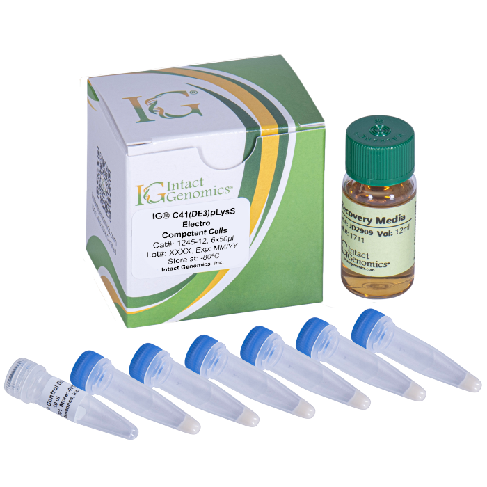
Intact Genomics (IG®) C41(DE3)pLysS Electrocompetent Cells are designed for transformation and routine recombinant protein expressed from a vector driven by an IPTG-inducible T7 promoter.
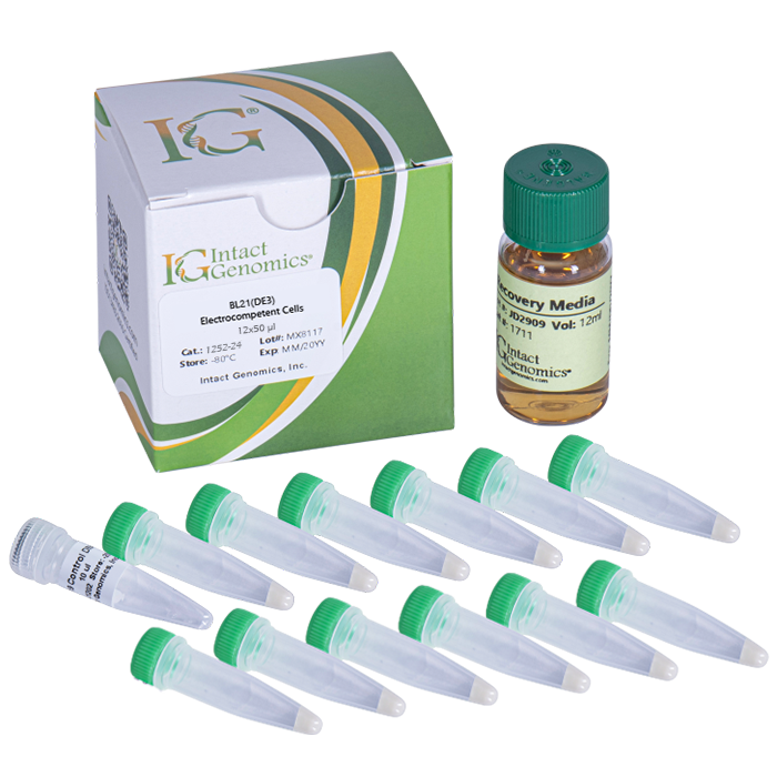
The C41(DE3) strain is a derivative of BL21(DE3) , was selected as survivor cells over-expressing the toxic oxoglutarate-malate carrier protein (OGCP), has lacI and other mutations, which prevent cell death associated with expression of many other recombinant toxic proteins. As a result, the C41(DE3)pLysS Electrocompetent Cells is highly effective in expressing toxic proteins from all organisms, including viruses, bacteria, yeasts, animals, plants, and mammals.
In addition, C41(DE3)pLysS strain also carries a chloramphenicol-resistant plasmid that encodes T7 lysozyme, which is a natural inhibitor of T7 RNA polymerase. Cells containing pLysS produce a small amount of T7 lysozyme. This strain is used to suppress basal expression of T7 RNA polymerase prior to induction, thus stabilizing recombinants encoding particularly toxic proteins. The effectiveness of C41(DE3)pLysS strain for expressing toxic proteins has been validated in >400 publications including all mutations were unveiled in genome sequencing.
Specifications:
Competent cell type: Electro competent
Species: E. coli
Strain: C41(DE3)pLysS
Format: Tubes
Transformation efficiency:≥ 1.0 x 10⁹ cfu/µg pUC19 DNA
Blue/white screening: No
Shipping condition: Dry ice
Reagents Needed for One Reaction
IG® C41(DE3)pLysS Electrocompetent cells: 25 µl
DNA (or pUC19 Control, 10 pg/µl): 1 µl
Recovery medium: 1 ml
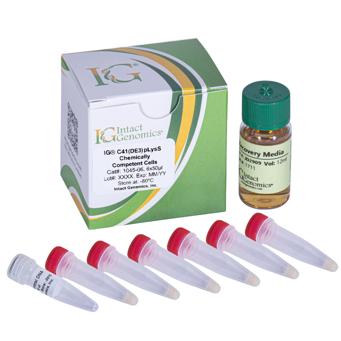
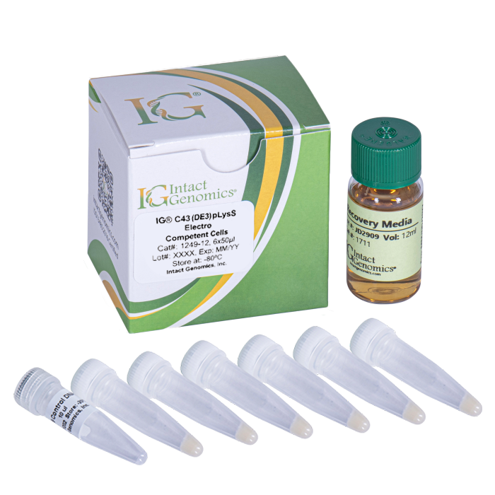
Product Components and Recommended Storage Condition:
- IG® C41pLysS(DE3) Electrocompetent Cells: -80°C
- DNA (pUC19, 10 pg/µl): -20°C
- Recovery medium: 4°C
Genomic Features:
IG® C41(DE3)pLysS Electrocompetent Cells have the following features:
- T7 Expression Strain
- Selected for expression of toxic proteins
- Suitable for expression of toxic genes
Quality Control:
Transformation efficiency is tested by using the pUC19 control DNA supplied with the kit and the high efficiency transformation protocol listed below. Transformation efficiency should be ≥ 1.0 x 10⁹CFU/µg pUC19 DNA.
Untransformed cells are tested for appropriate antibiotic sensitivity.
General Guidelines:
Follow these guidelines when using IG C41(DE3)pLysS Electrocompetent Cells.
- Handle competent cells gently as they are highly sensitive to changes in temperature or mechanical lysis caused by pipetting.
- Thaw competent cells on ice, and transform cells immediately following thawing. After adding DNA, mix by tapping the tube gently. Do not mix cells by pipetting or vortexing.
1245 1245-12 1245-48
Transformation Efficiency Calculation:
Transformation Efficiency (TE) is defined as the number of colony forming units (cfu) produced by transforming 1µg of plasmid into a given volume of competent cells.
TE = Colonies/µg/Dilution
Transform 1 µl of (10 pg/µl) pUC19 control plasmid into 50 µl of cells, add 950 µl of Recovery Medium. Dilute 10 µl of this in 990 µl of Recovery Medium and plate 50 µl. Count the colonies on the plate the next day. If you count 100 colonies, the TE is calculated as follows:
Colonies = 100
µg of DNA = 0.00001
Dilution = 50/1000 x 10/1000 = 0.0005
TE = 100/.00001/.0005 = 2.0×10¹⁰
High-Efficiency Transformation Protocol:
Use this procedure to transform IG® C41(DE3)pLysS Electrocompetent Cells. Do not use these cells for chemical transformation.
1. DNA/Cell Mixture Preparation
1.1. Remove competent cells from the –80 °C freezer and thaw completely on wet ice (10–15 minutes). Place sterile electroporation cuvettes and microcentrifuge tubes on ice. Bring IG Recovery Medium to room temperature.
1.2. Aliquot 1–5 µl DNA (1 pg–100 ng) into chilled microcentrifuge tubes on ice. If using the pUC19 control, add 1 µl pUC19 DNA (10 pg/µl) to a chilled microcentrifuge tube.
1.3. Once the cells are thawed, gently add 25 µl of competent cells to each DNA tube while keeping the mixture on ice. Mix gently by tapping the tube 4–5 times.
Do not pipette up and down or vortex, as this may damage the cells and reduce transformation efficiency.
2. Electroporation
2.1. Pipette 26 µl of the cell/DNA mixture into a chilled electroporation cuvette, avoiding bubbles. Quickly flick the cuvette downward to ensure the mixture settles evenly across the bottom of the cuvette, then proceed with electroporation.
Standard electroporation settings for E. coli:
Voltage: 1.8 kV, Resistance: 200 Ω, Capacitance: 25 µF, Cuvette gap: 0.1 cm.
3. Cell Recovery
3.1. Immediately add 974 µl of room-temperature IG Recovery Medium (or another suitable medium) to the cuvette. Gently pipette up and down three times to resuspend the cells.
3.2. Transfer the entire mixture to a culture tube (17 mm × 100 mm) and incubate in a 37 °C shaking incubator at 210 rpm for 1 hour.
4. Cell Plating
4.1. Prepare pre-warmed selection plates during the recovery period. Plates should be equilibrated to 37 °C and free of condensation to prevent contamination and mixed colonies.
4.2. Dilute the recovered cells 10–100X, if necessary, to obtain well-isolated colonies. Plate 20–200 µl of cells onto pre-warmed LB agar plates containing the appropriate antibiotic(s). For the pUC19 control, plate 50 µl of a 100X diluted transformation onto an LB plate containing 100 µg/ml ampicillin. Spread the cells evenly using a sterile spreader or autoclaved ColiRoller™ plating beads.
4.3. Incubate the plates overnight (12–16 hours) at 37 °C.
References:
- Walker JE & Miroux B. US6361966.
- Miroux B, Walker JE. J Mol Biol. 1996 260:289-98.
- Dumon-Seignovert L, Cariot G, Vuillard L.Protein Expr Purif. 2004 37:203-6.
- Wagner S, et al. Proc Natl Acad Sci U S A. 2008. PMID: 18796603
- Kwon et. al. Sci Rep. 2015 5:16076
- Zhang et. al. Microb Cell Fact. 2015 14:142.
- Bajzert et. al. BMC Vet Res. 2022 18(1):409.