
Sub-Total: $0.00
mVenus Fluorescence Standard Kit
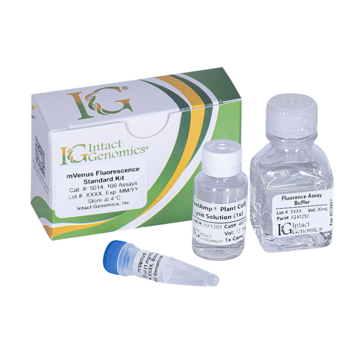
IG® mVenus Fluorescence Standard Kit contains purified recombinant mVenus protein for reliable quantification of mVenus and related yellow fluorescent protein signals. It supports standard curve generation, instrument calibration, and consistent fluorescence measurements using microplate readers, spectrofluorometers, confocal microscopes, and other compatible fluorescence-detection systems.
Custom formats, package sizes and bulk pricing are available upon request.
$486.00
Related products
-

TEV Protease
Price range: $115.00 through $1,875.00 Select options This product has multiple variants. The options may be chosen on the product page -
DsRed Fluorescence Standard Kit
$485.00 Select options This product has multiple variants. The options may be chosen on the product page -
Biotin Protein Ligase
Price range: $145.00 through $485.00 Select options This product has multiple variants. The options may be chosen on the product page


IG® mVenus Fluorescence Standard Kit contains purified recombinant mVenus protein for standardizing and quantifying GFP/YFP fluorescence in biological samples. mVenus is a bright yellow fluorescent protein (YFP) widely used in live-cell imaging and other fluorescence-based applications because of its strong fluorescence and high quantum yield.
Following the kit procedure, the amount of mVenus in a sample can be determined by comparing its fluorescence intensity with a standard curve generated from known concentrations of recombinant mVenus. The kit is suitable for fluorescence plate readers, spectrofluorometers, confocal microscopes, and other compatible fluorescence-detection instruments.
With a standard fluorescence plate reader, the IG® mVenus Fluorescence Standard Kit provides a detection limit of approximately 100 ng/mL. A lower detection limit may be achieved with higher-sensitivity instruments, such as spectrofluorometers or confocal microscopes. The excellent thermal stability of mVenus also makes the standard suitable for a broad range of fluorescence assays, instrument calibration, assay development, and quality-control applications.
Applications:
- Quantification of mVenus and YFP fluorescence in biological samples
- Standardization of fluorescence from mVenus -transfected cells using FastAmp® Plant Cell Lysis Solution
- Calibration of spectrofluorometers and other fluorescence instruments for in vitro mVenus quantification
- Generation of recombinant mVenus standard curves
- Use as a positive control in fluorescence assays
Benefits:
- Enables reliable and reproducible mVenus/YFP quantification
- Supports instrument calibration and assay optimization
- Compatible with multiple fluorescence-detection platforms
- Provides a detection limit of approximately 100 ng/mL with standard fluorescence plate readers
- Thermostable and convenient for routine fluorescence assays
Product Source:
Recombinant mVenus expressed from a plasmid carrying the thermostable YFP gene.
Shipping Conditions:
Ship with ice packs
Specifications:
- Purified recombinant mVenus protein
- Molecular weight: 31 kDa per monomer
- Free of detectable endonuclease activity
Components and Storage Temperature:
The following reagents are supplied with this product:

Quality Control:
Protein Concentration: The stock protein concentration is determined by UV absorbance and confirmed by running against a standard using PAGE densitometry.
Fluorescence Quantum Yield: Protein dilutions and fluorescence measured 515 nm excitation and 530 nm emission match a mVenus batch control standard.
References:
- Kremers, G.-J., Goedhart, J., van Munster, E. B. & Gadella, T. W. J. Cyan and Yellow Super Fluorescent Proteins with Improved Brightness, Protein Folding, and FRET Förster Radius,. Biochemistry 45, 6570–6580 (2006).
Preparation for mVenus Standard Curve:
This protocol is designed for use with a fluorescence plate reader. Sample volumes may be adjusted as needed for cuvettes or microscope specimen wells.
NOTE: A user may their own sample buffer as needed. The series below uses the kit’s assay buffer.
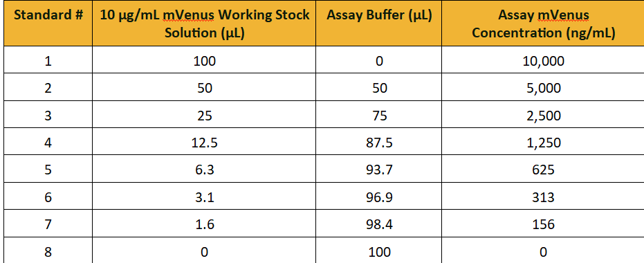
- Prepare a 1 to 100 dilution of mVenus into Fluorescence Assay Buffer to make a 10 µg/mL working stock solution
- Set up a dilution series into vials using the table below as a reference.
- Briefly mix the standards and measure the fluorescence amplitude of each sample. Set the excitation to 550 nm and the emission to 583 nm for mVenus standard curve measurements.
Tissue Preparation with FastAmp® Plant Cell Lysis Solution (Optional):
- Add 500 µL of 1X FastAmp® Plant Cell Lysis Solution to 50 mg of tissue in a 1.5 mL microcentrifuge tube. Prepare a blank control containing lysis solution without tissue.
- Manually grind tissue with a micro-pestle for 30 seconds.
- Centrifuge the samples in a benchtop centrifuge at maximum speed for 5 minutes.
- Carefully transfer tissue extract supernatant to fresh 1.5 mL conical tubes
- Transfer 100 µL for fluorescence intensity measurements using the same instrument settings used for the standard curve.
- Compare the fluorescence of the sample blank with that of the standard-curve blank for background fluorescence.
Plot mVenus Standard Curve:
- Plot fluorescence intensity (RFU) as a function of the Assay’s mVenus Concentration provided in the table above. It is appropriate to subtract the background buffer from all standards and samples.
- Fit the data points using linear regression, i.e. a line, to connect all data points. A good fitted curve will intersect all mVenus standard data points. The equation will have the form below:
RFU = slope x [mVenus] + (y-intercept) - Use the equation of the line to interpolate the sample’s concentration of mVenus:
[mVenus] sample = (RFU – y-intercept)/slope
mVenus Fluorescence Standard Kit Manual
DOWNLOAD
mVenus Fluorescence Standard Kit MSDS
DOWNLOAD
Certificate of Origin – mVenus Fluorescence Standard Kit
DOWNLOAD
"*" indicates required fields